Programs Prostate
Prostate Imaging
Optimizing imaging utilization and enhancing the quality of prostate imaging
What is the MUSIC Imaging Initiative?
- Goal: Improve radiographic staging of men with prostate cancer.
PSMA PET:
Prostate-Specific Membrane Antigen Positron Emission Tomography (PSMA PET) is an advanced imaging technique used to detect prostate cancer. It combines PET scanning with a tracer that binds to PSMA, a protein commonly found on prostate cancer cells. PSMA PET is highly sensitive and can identify cancer that might be missed by other imaging methods, making it a valuable tool for diagnosing, staging, and monitoring prostate cancer.
Current uses of PSMA PET:
- Diagnosis and Staging
- Detection of Recurrence
- Treatment Planning
- Monitoring Response to Treatment
- Clinical Trials and Research
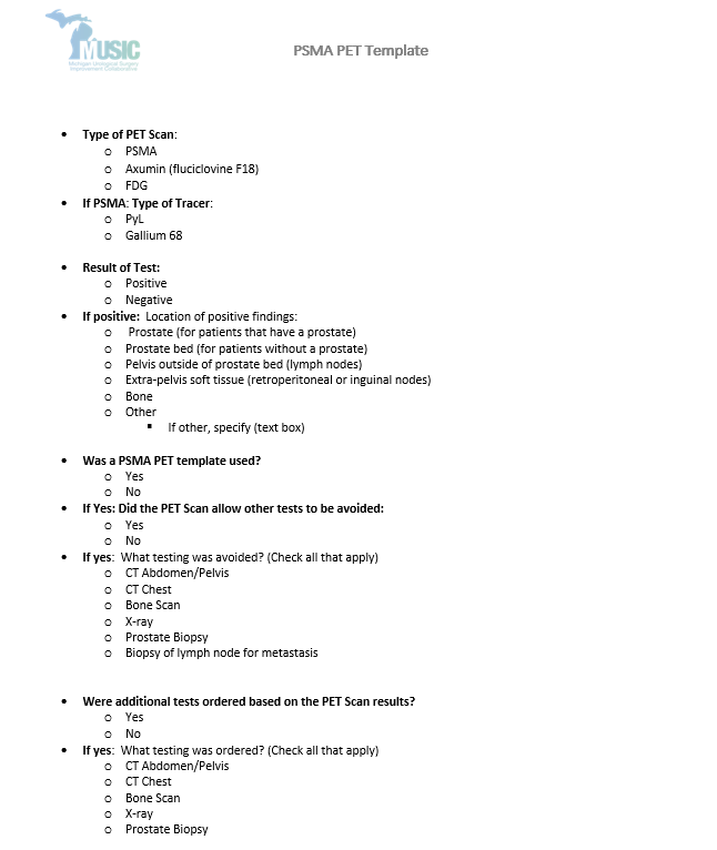
MUSIC started collecting PSMA PET variables in 2023 in hopes of:
- Developing PSMA PET appropriateness criteria.
- Reducing redundant imaging for patients with prostate cancer.
- Understanding how PSMA PET influences treatment decisions.
- Understanding the role of PLND in patients with prior PSMA PET scans.
Early work:
- Improving the quality of prostate MRI and fusion biopsies throughout Michigan.
- Optimizing imaging utilization for patients with newly diagnosed prostate cancer.
- To better understand the current rates of utilization and ensure that imaging studies were being ordered for patients that could truly benefit.
- MUSIC was able to achieve a statewide decrease in the utilization of both bone and CT scans for patients with low-risk prostate cancer.
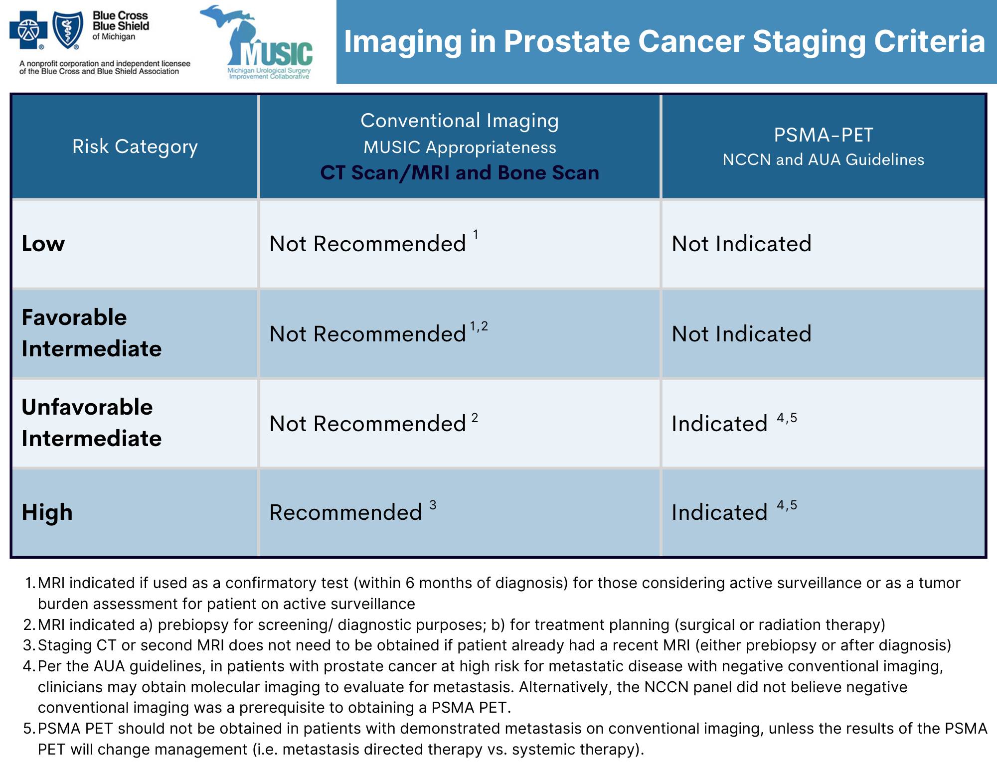
- To optimize imaging in intermediate/high-risk patients, MUSIC developed and implemented statewide specific, evidence-based appropriateness criteria for staging bone scan and/or CT scan (see below).


